It is well known that x-rays used for surgical planning vary in magnification. This variation exists in part because of the diversity of techniques used by x-ray technologists, patient positioning, and the patient’s size. Even with strict imaging protocols, the magnification of an x-ray image can vary. A thin patient lying on an imaging receptor is only a few inches above the plate, giving little distance for the X-rays to diverge. In a larger patient, the distance may be twice as far from the plate, allowing the divergent beam to spread much more before it hits the plate. This can result in a magnification factor of approximately 20%. Scaling an x-ray image is mandatory to assure the highest level of accuracy throughout the planning process, especially orthopedic templating. Does size matter when it comes to magnification of an x-ray and surgical planning? Absolutely! Preoperative planning aims to reduce the risk of intraoperative fracture and decrease overall surgical time. Images that are used for surgical planning must be accurately scaled to allow for accurate implant selection.
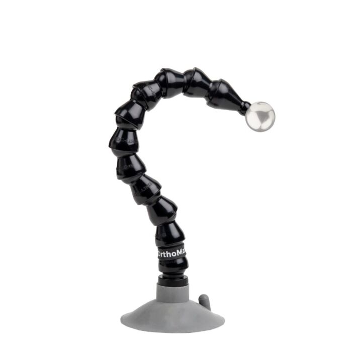
OrthoMark® - OM-1006 Short Suction Base Surgical Planning Marker
- SKU
- OM-1006
Description / OrthoMark® - OM-1006 Short Suction Base Surgical Planning Marker
OrthoMark was designed over 20 years ago to help measure x-ray magnification in digital images and they’ve been doing the job since. OrthoMark is compatible with all medical, orthopedic and veterinary templating software available, and there is a solution for every x-ray room.
The “shorty 2.5” quick suction cup Model OM-1006 is designed for use on smooth surfaces, by fast techs. When using, prepare OrthoMark for it’s destination by getting it as close to the desired plane of anatomy as possible. Then, holding the base in one hand, the end of the arm in the other, and in one deft movement, plunge the suction cup downward and simultaneously finely position the sphere. This should offer about two to five minutes of holding power. When finished, it is a good idea to lightly clean the surface with water and place on a counter or in a cabinet to avoid dust gathering. It is also best not to leave the suction cup attached to a wall or your equipment. The suction will eventually dissipate, and the unit will fall.
Position the sphere normally after positioning patient. By positioning OrthoMark's solid, 1 inch (25.4mm) stainless steel calibration ball at the precise anatomical plane of the shoulder, hip, knee—or any anatomy for that matter—during x-ray imaging, surgical planning software is able automatically find the x-ray marker in the image, calculate the x-ray magnification factor, and determine the true size of the anatomy in question. This helps to ensure that the appropriate prosthetic is selected before surgery.
The OM-1006 can be used with all orthopedic and veterinary surgical templating software.
Specifications
- Arm length: 10″
- Arm Weight - 3 oz
- Total Weight: 8oz
- Minimum Radius - 4.5"
- Ball Type - Stainless 316
- Ball: 25.4mm/1″ Stainless
- Base Type – 2” suction for smooth surfaces
OrthoMark® is available with a variety of bases and attachment methods and comes with a 25" arm which can be easily extended at any time. All parts are US supplied and readily available. The stainless-steel calibration ball is solid and doesn't degrade when surrounded by larger body mass.
More Information
| SKU | OM-1006 |
|---|---|
| Shipping Details | 6 x 5 x 3 @ 2 lbs |
| Shipping Method | UPS |
| Warranty | 1 year for Manufacturer Defects |
| Condition | New |
| Special Price | $150.00 |