Radiology Medical Imaging Stretchers for 2023
Z&Z Medical offers a wide variety of stretchers with unique features and designs which makes shopping for the stretcher to meet your specific needs easy! Below are a list of the most commonly requested stretchers for Imaging:
General Transport Stretcher – a basic stretcher for patient transport. Easy to use and priced well for most budgets.
Bariatric Stretchers are optimal for bariatric patients up to 1000 lbs. This stretcher can even fit through standard doors and passageways.
Hydraulic Stretchers are versatile and commonly used in transport, procedure, and recovery settings. With sturdy hydraulic elements, you can easily change the height with the push of a simple foot pedal. Designed with a shortened base for positioning in small rooms with large imaging equipment.
All Purpose Electric Powered Stretchers Versatile, easy to use, and time-efficient. Treat, transfer, and transport patients on a comfortable chair configuration. Provides positioning capabilities for a variety of procedures and all phases of treatment. Pre-induction, transport, treatment, and recovery can be performed on the same unit to improve case turnaround and streamline patient handling. Reduces staff time and prevents staff injury involved in transferring patient during various phases of care.
Multi Purpose Stretchers can be used as a stretcher, a chair, and even an exam/treatment table. This stretcher performs a lot of functions while taking up very little space and comes with a wide variety of optional accessories for a wide variety of imaging and medical procedures. Ideal for ER, PACU, OR, and L&D departments. Its multiple uses help eliminate patient transfers providing cost and time savings and enhanced safety for both patients and staff.
MRI Stretchers and Compact Adjustable Trolleys are designed to take patients in and out of MRI suites safely. MR safe.
Trauma Stretchers is intended for transport, treatment, and recovery of patients’ intra-and inter-departmentally in healthcare facilities. This unit offers unparalleled full-length X-ray capabilities, a treatment table and easy-rolling casters to provide full mobility.
Fluoroscopy Stretcher is designed for use with C-Arms for fluoroscopy-guided procedures. The unique design, with a proprietary sliding radiolucent top, enables a broader range of imaging capacity without repositioning the patient. The hourglass-shaped trim base accommodates a wide range of C-Arms. The security of a specialty imaging table with the mobility of a stretcher. It’s full-length radiolucent top is perfect for surgery, interventional, and pain management procedures.
Take a few moments to browse the wide selection of radiology imaging and medical stretchers that we have to offer at www.zzmedical.com
Chiropractic Digital X-Ray Upgrade - "I wish I had done it years ago"
Z&Z Medical recently sold and installed a New Digital X-ray Unit with DR from 20/20 Imaging with OpalRad software at Eberline Family Chiropractic and Acupuncture in Grundy Center, Iowa. While completing the calibration to the unit, Dr Eberline stopped by and thanked us for finally getting him to go digital! He said, “It’s already been well worth the money I’ve spent and the best part of upgrading to digital X ray is the resulting improvement in the quality of care I can give my patients. I am able to get better images more quickly for my patients. I wish I had done it years ago. We have taken more images so far in 2021 than we did all of last year.”
We at Z&Z Medical often hear hesitation from our customers to convert to digital for financial reasons. It is most often the case that months later we hear that they wish they had done it sooner. As with Dr Eberline, most of them admit that they take far more x-rays now then when they had analog imaging. The time saving over analog makes it far easier and more convenient to take an x-ray if they have any hesitation about the patients physical condition. Plus the image quality is always sharp and very dependable, as opposed to film and old chemicals.
If you still have an analog system and would like to learn more about the benefits of digital and speak to facilities that have made the leap – give us a call. We would love to share with you many success stories and add you to the growing list!
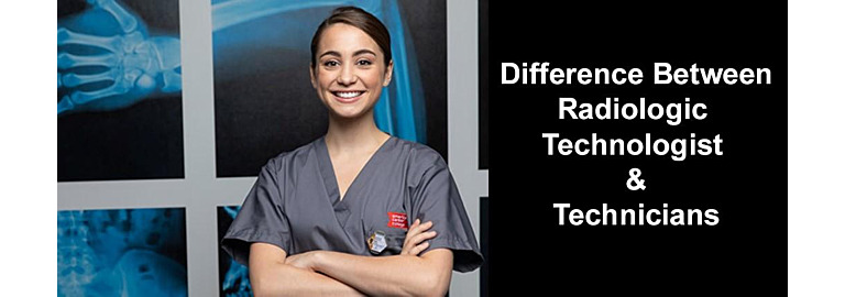
The Difference Between Radiologic Technologists and Radiologic Technicians
Radiologic technologists and technicians both work in the field of Radiology, but the training and responsibilities are very different. Both professions involve working with patients and performing an imaging procedure, but technologists have more education and take on more responsibilities than radiologic technicians do.
Radiologic Technologist
Radiologic technologists are expected to earn an Associate degree at a two-year program from an accredited educational institution. Many accredited programs require certain pre-requisite college classes to be completed before even applying to the radiology program. Technologists are licensed by the ARRT (American Registry of Radiologic Technologists.) as well as licensed in the state in which they work
Digital X-Ray Viewing for Tight Spaces
In most Chiropractic and small clinic offices floor space is a premium luxury. To keep monthly rent and lease payments affordable you have to squeeze as many services as possible into the smallest space in your office. Many times, you are forced to get double duty out of one room. Adjustment or procedure rooms get used as dressing or waiting areas while office and clerical duties are performed in kitchens and breakrooms. This is especially true when it comes to the x-ray room in many practices. Since x-rays are not performed continuously throughout the day, these rooms are typically small and quite often used for other services such as massage, adjustments, and procedures. In the days of film developing, this was further complicated by the need for a darkroom. Most x-ray rooms had a darkroom located adjacent to or down the hall from x-ray. You had to leave the patient unattended while you went to develop a series of x-rays. This also took up valuable floor space.
Size Does Matter: X-Ray Image Scaling Devices
It is well known that x-rays used for surgical planning vary in magnification. This variation exists in part because of the diversity of techniques used by x-ray technologists, patient positioning, and the patient’s size. Even with strict imaging protocols, the magnification of an x-ray image can vary. A thin patient lying on an imaging receptor is only a few inches above the plate, giving little distance for the X-rays to diverge. In a larger patient, the distance may be twice as far from the plate, allowing the divergent beam to spread much more before it hits the plate. This can result in a magnification factor of approximately 20%. Scaling an x-ray image is mandatory to assure the highest level of accuracy throughout the planning process, especially orthopedic templating. Does size matter when it comes to magnification of an x-ray and surgical planning? Absolutely! Preoperative planning aims to reduce the risk of intraoperative fracture and decrease overall surgical time. Images that are used for surgical planning must be accurately scaled to allow for accurate implant selection.